
Neurosurgeons often face tough choices when operating to remove a tumor from a patient’s brain. If they leave bits of the tumor behind it’s likely to regrow, but cut away too aggressively and the patient could be seriously impaired.
New technology developed by a University of New Mexico Health Sciences researcher promises to provide neurosurgeons with critical real-time information about the tumor and surrounding cortical tissue, enabling better outcomes.
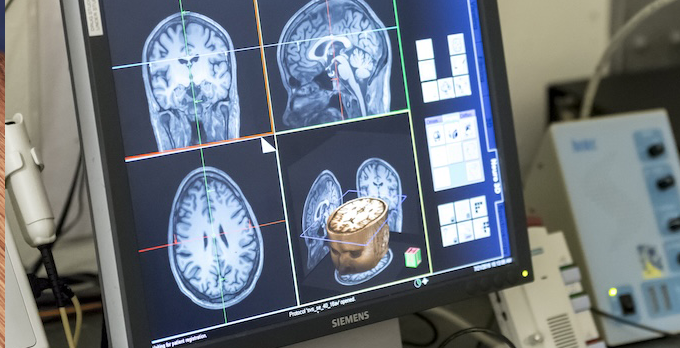
Stefan Posse, PhD, a professor in the UNM Department of Neurology, with a secondary appointment in Physics & Astronomy, has received a patent for data acquisition and analysis software that enables operating room magnetic resonance imaging (MRI) machines to better target active tumors while identifying adjacent brain tissue with important sensory, motor and language functions that should be spared.

It provides a wealth of functional and metabolic information in a single scan, and it’s very compatible with existing clinical MRI scanners. We can get a clinically meaningful scan on a patient with a brain tumor in just three minutes.
“The interesting thing about this is that it provides a wealth of functional and metabolic information in a single scan, and it’s very compatible with existing clinical MRI scanners,” Posse said. “We can get a clinically meaningful scan on a patient with a brain tumor in just three minutes.”
MRI scanning relies on the ubiquity of hydrogen in living tissue (water – H2O – is the most abundant molecule in the body) and the fact that the nucleus of a hydrogen atom contains a single proton. MRI machines create a powerful magnetic field that, coupled with radio waves, briefly polarize the protons, causing them to emit radio signals that can be processed into an image, Posse said.
Structural MRI scans create a picture reflecting different densities of tissue and signal relaxation properties in the body, such as the brain.
Another MRI method – functional MRI (fMRI) – highlights tissues that are metabolically active. In fMRI brain imaging, the scanner detects the heightened blood flow that occurs when a brain network becomes active, Posse said. In addition, fMRI can also detect the subtler activity of functional networks even when the brain is resting.
MRI can also be used for spectroscopic imaging, which identifies organic molecules that are unique to a specific type of brain tumor, helping radiation oncologists to direct their treatment efforts. “A recent study using this biochemical information to guide radiation oncologists to direct their treatment efforts to active tumors showed improved survival of patients,” he said.
Neurosurgeons can use functional and spectroscopic MRI scans to guide them as they remove cancerous tissue while trying to avoid damaging nearby tissue needed for normal neurological functioning, Posse said. But both of these methods are time consuming, with each scan usually being conducted in separate sessions.
Posse’s patent represents a new way of programming MRI scanners so that they can perform both tasks at once with the help of a sophisticated data analysis tool.
“The goal for this particular patent is going beyond the current paradigm of collecting one type of data at a time by collecting multiple image modalities at the same time,” he said. “We are combining functional MRI with metabolic imaging – spectroscopic imaging. It significantly shortens the overall scan time. We have a very powerful way of collecting data very rapidly with our high-speed functional and spectroscopic imaging technique.”
Currently, neurosurgery is often performed on patients who are awake and answering questions or performing a task so as to determine whether the next cut will damage a critical structure, Posse said. Combining functional and spectroscopic MRI scanning will provide neurosurgeons with better information and provide for the patient to be anesthetized.
“Neurosurgeons have always been focused mostly on the motor system, the language system and maybe the visual and auditory sensory system,” he said. “But the frontal cortex has always been an area where they don’t have a lot of leverage.
“This is where resting state MRI comes in. It allows us to map the brain comprehensively in all areas – the frontal cortex in particular, which is not feasible with conventional task-based functional MRI brain scanning. If you collect enough data, you can dissociate up to 100 different resting state networks that represent different functions of the brain.
Preliminary results from a study led by Posse and collaborators at the University of Minnesota and University of Pittsburgh show that resting state MRI works even in anesthetized patients. “You can have the patient under anesthesia, and you can still see resting state networks,” he said. “That opens up a lot of opportunities in the way patients are being treated.”
Posse has recently received a sizable Small Business Technology Transfer grant from the National Institutes of Health to further develop his innovative real-time resting-state fMRI brain imaging methodology. His research lies at the intersection of physics and modern medicine, but his foremost concern is about how his work will improve patient outcomes in a real-world clinical setting.
“The goal is to integrate this into patient care,” Posse said, “and our goal really is to make a difference in care and surgical outcomes for patients with brain tumors.”