Reflectance confocal microscopy (RCM) is an exciting new imaging tool that helps clinicians in monitoring and diagnosis of skin lesions, including skin cancer. Imagine being able to see deeper into the skin without cutting. This new imaging modality can empower providers to diagnose skin cancer in their patients without putting them through an unpleasant and scarring procedure, especially when the diagnosis is uncertain. RCM is painless, non-invasive, and well-liked by patients of all ages. Here at UNM, we are specifically studying the use of this imaging tool in children with skin conditions.
Assistant Professor John Durkin, MD has expertise in reflectance confocal microscopy and pigmented lesions, and is the only such specialist in the state of New Mexico. He enjoys educating physicians on the principles of confocal microscopy and how they can integrate it into their clinical practice. He also actively trains residents and medical students on the basics of confocal image interpretation which is a unique attribute of our program.
How is reflectance confocal microscopy used?
When a provider identifies a lesion of concern, the area is marked and photographed.
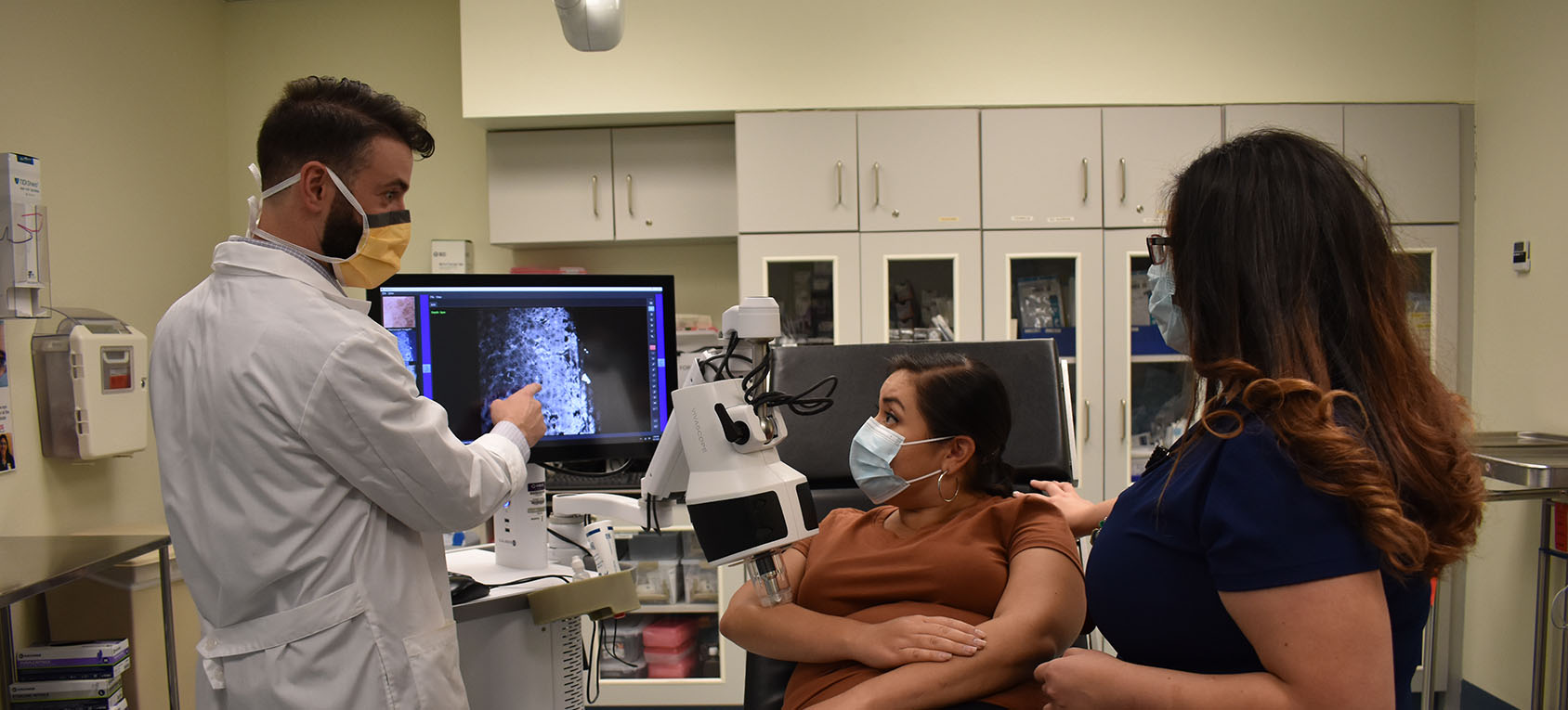
Our trained imaging technician captures images using the confocal microscope in a painless procedure that takes approximately 15 minutes.
The images are then interpreted by our expert and a report is sent to the ordering provider who will decide if any further treatment or removal is needed or if the area needs to be reimaged in the future.
This procedure can therefore essentially take the place of an invasive biopsy in many situations.